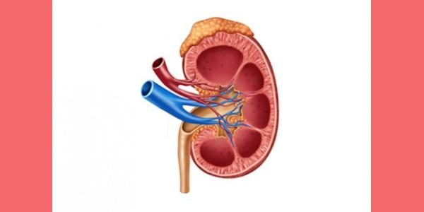
- 1. N. B. age: 60 years. Her son reported to chamber on 07 April 2015. She was admitted in a renowned hospital undergoing dialysis of kidneys three times in a week. Creatinine – 6+. Both the kidneys are affected. Right kidney fully damaged, Left one almost damaged.
- CT Scan of whole abdomen done on 25 September 2014. (Attached As Annexure- A).
Right kidney:
- Non contrast CT scan showed increased attenuation through all perirenal fat and slightly masked by fine, dense radial structures. Perirenal space is broadened, possibly as sequel of inflammatory change with formation of granulation tissue and accumulation of exudate, associated with thickening of the adjacent peritoneum.
- Complete loss of normal architecture of right kidney. Possible distorted parenchyma with exudates; degraded blood and urine are seen at the site of right kidney.
Left kidney :
Thinning of left renal parenchyma. No evidence of hydronephrosis, cyst, calculus or any other mass lesion.
Impression of CT scan. Features are suggestive of massive right renal inflammation with maceration of right renal parenchyma with accumulation of perirenal urine/ exudates/ degraded blood. D/D less likely neoplasm. Advised other adjuvant examination (may include CT guided FNAC) for further evaluation. Thinning of left kidney renal parenchyma, possibly sequel of CKD. Mild hepatomegaly. Calculus cholecystitis.
- The Ultrasound taken for the same patient on 29 September 2014. (Attached As Annexure- B).
Report shows the following:
Right kidney: Right kidney is enlarged in size. Large mixed echogenic mass with echogenic strands measuring about 13x11cm in size are seen in upper part of right kidney. No evidence of calculus could be seen. Pelvicaliceal systems of lower part of kidney are not dilated. Ureter is not dilated.
Left kidney: Left kidney is slightly small in size with ill-defined cortex and medulla. Bipolar length of left kidney measured about 85 mm. Cortical echotexture is raised in kidney. No evidence of calculus could be seen. Pelvicaliceal systems are not dilated. Ureter is not dilated.
Comment : 1) Suggestive of soft tissue mass with organizing haematoma in right kidney.
2) Chronic parenchymal disease of left kidney.
3) Mild hepatomegaly.
4) Suggestive of Cholelithiasis
5) Suggestive of small left kidney with bilateral renal
parenchymal disease.
- The case was recorded in homeopathic manner and the important aspects of the case were the following :
- Strong fear of death, dark and ghosts . She says to everyone, “Possibly I
would not survive”.
- Very fastidious, everything must be in order, her plate and glasses separate. Tension++. Frequent change into fresh cloths. Strict minded = wants the work to be done on time. Cannot reply on the face. Short memory – cannot remember names.
Always wiping her mouth. Cannot stay without taking a bath. Quite often rubbing hands and palms while sitting. Likes to get her body massaged. Heat of head. Cold washing amelioates. Urine scanty. Cold, constipation, pain in extremities amel by pressure. Sweating of head and shoulder, wetting the pillow during sleep. Coughing as soon as she drinks water. Likes warm food. Drinks water in sips. Sleep disturbed by pain. Burning of skin. Vomiting tendency especially of fish and meat.
- Appendix-operated on. BP-high. Oedema of extremities. Father – died of heart attack. Mother –rheumatism. Brother – rheumatism
- Symptoms considered for Repestorijation
Fastidious
Anxious & restless
Fear of death
Fear of dark & ghosts.
Nausea seeing food.
Head – heat and by cold washing.
Perspiration head, wetting the pillow.
Feet cracked soles
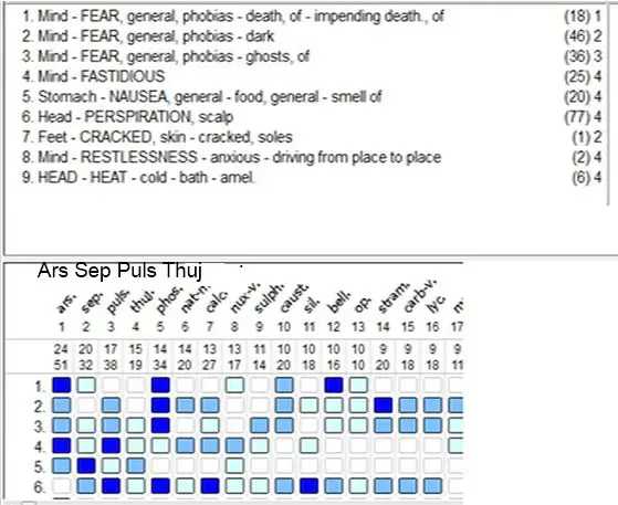
Rubrics considered :
- RM, Mind fear Gen Phobias, impending death of –Acon2,ARS3,asaf1, BELL3, bry1, caps1, carc2, Caust2,Croc2, cupr1, Lach1, MERC3, NV1, Op1, PHOS3, sep1, staph1.
- RM, Mind Fastidious : Alum, Arac2, ARS3,Ce2,CARC3, con1, graph2, lgn1, lod1, medo1, NM2, Ns2, NV2, phos1, PULS3, Sep1, Sil1, Sulph1, thuja1.
- RM, Mind Fear Phobias , Ghost of , Acon2 ,Ars2, CV2, caust2 , Hyos2, Kbr2 , lach3, lyc2, Manc2, PHOS3, plat2, puls2, stram2, sulph.
- RM, Mind fear Gen Phobias, Dark Acon2, Ars2, CC2 , cp1, cs1, camph2, CANNI3, Canns2, cavban2, Cv2, carc1,caust2, cham1, cupr2, gels1, grin2, lyc2, Manc2, Med2, Natm2, PHOS3,Puls2,STRAM3, strontc2.
- RM, Stomach Nausea Gen, food gen, smell of-aeth, Ars2 , ip2 , Nv1 , phos-ac1 , podo1 , petl1 , puls1 , SEP3 , stann1 , thuja2 , tab.
- RM, Head, perspiration scalp, agar2 , ANAC3 , Ant-t2 , Apis2 , Ars-i , Barc1 , Bar-m2 , Bell2 , CALC3 , cp2 , cs2 , cv2 , caust2 , CHAM3 , CHIN3 , graph2 , GUAJ3 , Hep2 , Kc2 , Kp2 , Lyc2, , Magm2 , MERC3 , Mez2 , MUR-AC3 , Ni-ac2 , Petr2 , PHOS3 , PULS3 , Pyrog2 , RHEUM3 , Sep2 , SIL3 , Stram2 ,.
- Feet, Skin Cracked, soles – Ars (single)
- RM, Mind Restlessness anxious driving from place to place – ARS3 , Tab.
- Synthesis 8.1, Head, Heat, cold bath- amel– Ars1 , Euphr1 , ind1 , Mez1 , Natm1 , sep1 .
Repertorization Result:
Prescription:
7/4/2015. 1. Ars Alb M/3, twice a day for 4 days, one tea spoonful from 2nd glass of water. Then gap for 4 days.
9/4/2015. Patient is in good mood and slept better. Nausea tendency much less than before. Appetite better.
15/ 4/2015. Thuja M/2, complimentary of Arsenic as well as augments the action of Ars, for 4 days+ 4 days gap.
24/4/2015- Improvements continues. Ars Alb M/3 for eight days.
2/4/2015- Improvement continues. As complimentary, anti sycotic and for augmenting the action of Arsenic, Medo M/2 for 8 days was prescribed.
10/4/2015. Improvement continues. The CT Scan was repeated. The result is very strange: The right kidney has come back to its normal shape and size. It was unbelievable to the allopathic doctors. Arsenic alb M/4. For 8 days was prescribed.
CT Scanning of whole abdomen was done on 05 May 2015. (Attached as Annexure- C).
The Impression was:
Kidneys: Right kidney is normal in size, shape and position. Measurement of right kidney is 11.3 cm.
Left kidney: Is smaller in size. Measurement of right kidney is 6.3 cm.
Cortical echogenecity is increased in both kidneys. Cortical- medullary differentiation is lost. Pelvicalyceal systems are not dilated.
19/4/2015. Improventment continues. Thuja M/3. For 8 Days.
28/4/2015. Improvement in all aspects continues. Creatinine level has reduced from 6+ to 3+.The patient was brought home, dialysis of kidney was being continued. Discontinued taking medicine.
COMPARISON OF REPORTS
| CT Scan Report on Sept 25, 2014 (before homeopathic remedy) | Ultra Sonography Report on 29 Sept 14(before homeopathic remedy) | CT Scan Repot – May 05, 2015.(after homeopathic remedy) | |
| Right kidney | Non contrast CT scan show, increased attenuation through all perirenal fat and slightly masked by fine, dense radial structures.
Perirenal space is broadened, possibly as sequel of inflammatory change with formation of granulation tissue & accumulation of exudates, associated with thickening of the adjacent peritoneum. Complete loss of normal architecture of right kidney. Possible distorted parenchyma with exudates, degraded blood & urine are seen at the right kidney. Impression: Features are suggestive of massive right renal inflammation with maceration of right renal parenchyma with accumulation of perirenal urine/ exudates/degraded blood. D/D Less likely neoplasm. |
Right kidney is enlarged.
Large mixed echogenic area with echogenic strands measured about 11×10 cm in size is seen in upper part of right kidney. No Evidence of Calculus. Pelvicaliceal systems of lower part of kidney are not dilated. |
Right kidney is normal in size, shape and position. Measurement of right kidney is11.3 cm & |
| Left Kidney | Left kidney:Thinning of left renal parenchyma. No evidence of hydronephrosis, cyst, calculus or any other mass lesion.
Impression: Thinning of left kidney renal parenchyma, possibly sequel of CKD. |
Left kidney is slightly small in size with ill defined cortex and medulla.
Bipolar length of left kidney measured about 85mm. Cortical echotexture is raised in kidney. No evidence of Calculus. Pelvicaliceal system are not dilated. Ureter isnot dilated. |
Left kidney: Is smaller in size. Measurement of right kidney is 6.3 cm. |
Annexure- A
|
|

HOUSE NO. 48, ROAD NO. 9/A, SATMASJID ROAD
DHANMONDI, DHAKA-1209
PHONE z 9126625-6, 9128835-7
|
E-mail 1 [email protected]
wwvv.ibnsinatrust.com
COMPUTED TOMOGRAPHY (128 SLICE)
- No. : D334683 Date : September 25, 2014
Patient’s Name : N.B. Part Scanned : Whole Abdomen
Age : 60 year(s) Sex : Female
Refd. by : Dr. Md. Abdus Salam. MBBS, BCS, FCPS, MRCS, MS.
CLINICAL INFORMATION:
- Pain at left right loin pain since 10 days.
- CKD 81 IHD.
TECHNIQUE:
10-mm contiguous spiral axial sections were obtained from hepatic dome to ischial tuberosities without oral, rectal & I/V contrast administration, and coronal & sagittal MPR images.
FINDINGS: *
- Liver : Mild hepatomegaly. It exhibits uniform parenchymal density. No
focal area with altered attenuation is seen in the liver. There is no
intrahepatic biliary duct dilatation. No fatty infiltration or fatty liver.
- IVC & PV : Normal.
- Spleen : Normal size & shape. No intraparenchyrnal focal or diffuse lesion is
noticed.
- GB : Multiple intraluminal radiopaque calculi with mild uniform thickening
of wall. No soft tissue lesion is seen within the lumen. CBD: Not dilated.
- Pancreas : No evidence of edema, cyst, calcification or focal parenchymal
lesion. Peripancreatic fat planes are preserved. Pancreatic duct is not dilated.
- Adrenals: Normal in size & position. No evidence of mass is seen at the site
of suprarenal glands.
- Right kidney:
- Non contrast CT scan show, increase attenuation of thorough all perirenal fat andslightly masked by fine, dense radial structures. Perirenal space is broadened, possibly as sequel of inflammatory change with formation of granulation tissue & accumulation of exudate, associated with thickening of the adjacent peritoneum.
- Complete loss normal architecture of right kidney. Possible distorted
parenchyma with exudate, degraded blood & urine are seen at the site of
right kidney.
- Left kidney: Thinning of left renal parenchyma. No evidence of
hydronephrosis, cyst, calculus or any other mass lesion.
- Ureters: No evidence of hyodro-ureter.
- Bladder: No intraluminal lesion or wall thickening is present.
- Uterus : Not bulky, and no altered attenuated mass is noticed.
- Bilateral adnexae: No parametrial/ovarian/tubual mass is evident.
- Mesentery 8: peritoneum: Attenuation appears to be normal.
- Enlargedsubdiaphragmatic, hepatic hilar, celio-mesenteric, paraaortic,
parailiac,
- Ascites : not present
- Right-sided pleural effusion.
Transcripted by Obayed U llah
HOUSE NO. 48, ROAD NO. 9/A, SATMASJID ROAD
|
|
DHANMONDI, DHAKA-1209
PHONE z 9126625-6, 9128835-7
E-mail 1 [email protected]
wwvv.ibnsinatrust.com
|
IMPRESSION:
- Features are suggestive of massive right renal inflammation with
maceration of right renal parenchyma with accumulation of perirenal
urine / exudate / degraded
blood. D/D. Less likely neoplasm. Adv. Other adjuvant examination (may include CT guided FNAC) for further evaluation, please. .
- Thinning of left kidney renal parenchyma, possibly sequel of CKD.
- Mild hepatomegaly.
- Calculus cholecystitis. Adv. Other adjuvant examinations for further
evaluation, please.
|
With compliments for referral,
- ABUL MOKARIM (Ex. Major)
MBBS (DU)., DTM (Nagasaki), Ph.D. (Japan).
Fellow-Resident, Nagasaki University Hospital,
Chief Consultant, Ibn Sina Diagnostic & Imaging Center
* This is only a professional opinion and not a diagnosis, hence it should be clinically correlated.
Annexure -B
|
|
Professor Dr. Md. Delwar Hossain
MBBS (Dhaka), DMRD (DU),
FCPS (Radiology & Imaging),
Specialist in Interventional Radiology,
CT scan & Ultrasound Imaging.
Professor & Head
NIKDU, Dhaka.
Ultrasonography Report
Name: N.B. Age: 60 Yrs. Date:29-Sep-14
Ref. By: Dr. Md. Abdus Salam MBBS, Bos (Health0,
FCPS (Surgery), MRCS, MS (Urology)
Name of Investigation: Ultrasonogram of KUB region.
Right kidney is enlarged in size. A
Large mixed echogenic mass with echogenic strands measured
about 13x11cm in size is seen in upper part of right kidney.
No evidence of calculus could be seen.
Pelviealiceal systems of lower part of kidney are not dilated.
Ureter is not dilated.
Left kidney is slightly small in size with ill-defined cortex and medulla.
Bipolar length of left kidney measured about 85 mm.
Cortical echotexture is raised in kidney.
No evidence of calculus could be seen.
Pelviealiceal systems are not dilated. Ureter is not dilated.
Urinary bladder is well filled and normal in contour. Wall thickness is normal.
|
Comment: 1.Suggestive of soft tissue mass with organizing
haematoma in right kidney.
- Chronic parenchymal disease of left kidney..
|
ANNEXURE- C
HOUSE NO. 48, ROAD NO. 9/A, SATMASJID ROAD
|
DHANMONDI, DHAKA-1209
PHONE z 9126625-6, 9128835-7
E-mail 1 [email protected]
wwvv.ibnsinatrust.com
| |
||
|
|
| ID NO : | R72229 | Scen Date : | May 05,2015 |
| Name of PT : | N. B. | Age : 61 year (s) | Sex : Femals |
| Referred by : | Ibn Sina Hospital | Bed/Cabin : | C440 |
| Part Scanned : | Whole Abdomen |
Whole Abdomen
LIVER : Mildly enlarged in size (15.0 cm). Parenchyma is homogeneous. No focal lesion is seen.
BILIARY DUCTS : Biliary tree is not dilated. C.B.D is of normal caliber.
GALL BLADDER : Gall bladder is normal in size. Wall thickness is within normal limit.Multiple echogenic structure casting posterior acoustic shadows are noted in the neck of the gallbladder.
PANCREAS: Pancreas is normal in size with normal echotexture. No focal lesion is seen
SPLEEN: Normal in size (7.2 cm) with uniform parenchymal echogenicity. No focal lesion is seen.
KIDNEYS : Right kidney is normal in size, shape and position. Left kidney is smaller in size. Measurement of right kidney is 11.3 cm & left kidney is 6.3 cm. Cortical echogenicity is increased in both kidney. Cortical- medullary
differentiation is lost. Pelvicalyceal systems are not dilated.
- BLADDER: is empty, so pelvic organs could not be properly evaluated.
UTERUS : Uterus is normal in size with homogenous myometrial echotexture.
Endometrium is normal in thickness. Cavity is central and empty.
ADNEXA : Both adnexal region appear normal.
CU-DE-SAC : No collection is seen. g
Others : No free fluid or any mass lesion is seen in the abdomino pelvic cavity.
|
|
HOUSE NO. 48, ROAD NO. 9/A, SATMASJID ROAD
DHANMONDI, DHAKA-1209
PHONE z 9126625-6, 9128835-7
E-mail 1 [email protected]
wwvv.ibnsinatrust.com
 |
||
| |
|
lMPRESSl0N 1. Mild hepatomegaly.
- Suggestive of Cholelithiasis.
- Suggestive of small left kidney with bilateral renal
parenchymal disease.
Adv : Please correlate with clinical & biochemical findings.
 |
|
Dr. Kamrun Nahar
M.B.B.S MD
Consultant Radiologlst
Ibn Slna Medical College Hospital.


Thank you Dr. Rahman for sharing your truly excellent case with our readers.
Alan V. Schmukler
Very nice case.Well explained.
Dr Rahaman … I am writing this from the USA … Could not understand what M/3 and M/2 was, in your excellent article … Could you please explain … Pradhan
e -mail ID :- [email protected]
in atrophy kidney plumbum is useful,terebinth30 i have noticed in chronic cases being prescribed by many homeopaths.addisons disease of course arsenic and nat mur commonly prescribed.reckeweg combinations for kidney very imp.