In nerve conduction tests, conductivity of the nerve is measured by placing two electrodes on the skin above the nerve, and the flow of current is measured between the two electrodes. The accepted belief in neurobiology is that nerve fiber conducts impulses electrically through depolarization and repolarization of the membrane. However this belief lacks a sound theoretical basis as nerves contains both water and ions, and in currently accepted science an aqueous solution is believed to conduct electrical energy through ions.
In the living being, nerve signals are believed to be transmitted by action potentials. The action potential being a sudden change in the membrane potential of nerve fiber from its normal resting negative potential to a positive potential and thereafter most rapidly returning back to its normal negative potential through the following stages:
1. A resting or polarized stage being the initial one, when the membrane potential is regarded to have negative value of about –90 millivolts .
2. In the depolarization stage the normal state of –90 millivolts in the interior of the fiber is neutralized due to a sudden increase in permeability of the membrane to sodium ions (Na+). This accounts for rising of potential in the positive direction and is termed depolarization.
No action potential however initiates when the nerve fiber membrane remains undisturbed. But when any disturbances occurs (mechanical disturbance, electrical impulses, chemicals, etc.) for which there is a sudden rise in membrane potential of 15 to 30 mV i.e. about -65 mV or above (called threshold potential), an action potential is said to begin.
Thus in general, for normal propagation of an impulse to occur the ratio of action potential to threshold for excitation , called the safety factor, must always be greater than unity. Though an action potential crops up at one spot on the membrane, it usually excites adjacent portions of the membrane and propagates in all directions away from the stimulus, until depolarization of the entire membrane is attained. Further, the spread of depolarization stops as soon as the action potential reaches a point on the membrane where it is unable to generate the threshold potential for stimulating the area of the membrane in its vicinity.
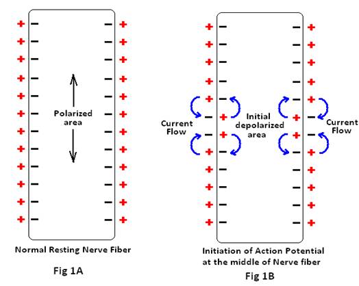
The mechanism of propagation of action potential follows. A normal resting fiber is shown in Fig 1A. When a nerve fiber is excited in the mid portion i.e. there is a sudden increase in the permeability of Na+ ions at the mid portion giving rise to an initial depolarized area (Fig 1B).
The positive electrical charges carried by the inward-diffusing sodium ions through the depolarized membrane propagate for several millimeters in both directions along the core of the axon. These positive charges often increase the voltage above the threshold level, up to a distance of some millimeters inside fiber, for initiating an action potential. Therefore, induced opening of the sodium channels in these new regions helps rapid spread of action potential in that region.
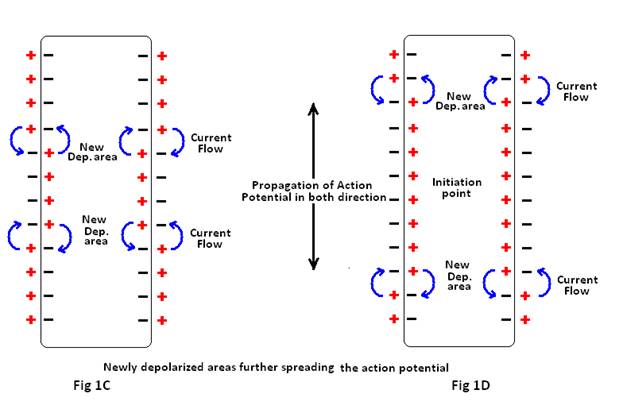
These newly depolarized areas further spread the action potential along the membrane by the similar mechanism, causing progressively more depolarization. Thus, the depolarization process travels along the entire length of the fiber (Fig 1C). This transmission of the depolarization process along a nerve or muscle fiber is termed a nerve or muscle impulse. It is believed that as long as the membrane is still depolarized due to the former action potential, a new action potential cannot crop up .This is due to the fact that shortly after the action potential is initiated, the sodium channels (or calcium channels, or both) become inactivated, and no amount of excitatory signal applied to these channels at this point will open the inactivation gates. The only condition that will allow them to reopen is for the membrane potential to return to near the original resting membrane potential level. Then, within another small fraction of a second, the inactivation gates of the channels open, and a new action potential can be initiated. The period during which a second action potential cannot be elicited, even with a strong stimulus, is called the absolute refractory period. This period for large myelinated nerve fibers is about 1/2500 second. Therefore, it is apparent that such a fiber can transmit a maximum of about 2500 impulses per second.
3. The repolarization stage is reached within 10-4 seconds of depolarization. Here, the sodium channels start closing and potassium channels open more than normal, resulting in rapid diffusion of K+ ions to the exterior and re-establishment of the normal negative resting membrane potential1.
To explain the conduction of electrical impulses in nerve fibers, the following experiments are performed :
Experiment 1: A circuit is prepared with an LED , connected to a 9 volt battery through distilled water and a human being (i.e; electrodes are held between forefinger and thumb) connected through graphite electrodes as shown in fig 2.
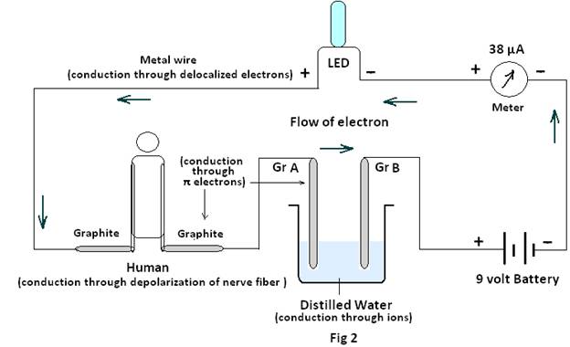
The LED is found to glow and the current recorded to be about 34 to 38µA. Considering the circuit to be divided in four parts. it is believed that i) for the metal part electrical energy is conducted by the metals through delocalization of electrons ii) for graphite it is conducted through π electrons and iii) for the humans it is conducted through nerves by depolarization & iv) for the distilled water it is conducted through ions.
Experiment 2: Two different circuits each prepared with an LED , a 9 volt battery and two graphite electrodes are placed on the skin of an arm such that current flows in the opposite direction through the circuits as shown in Fig3.
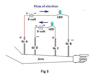
Observations: Both the LED’s glow.
Experiment 3: This used two different circuits each prepared with an LED , a 9 volt battery having a common positive graphite electrode (Gr A) and separate negative electrodes Gr B and Gr C as shown in Figure4.The condition of the LEDs are noted for the following situations;
I) When only Gr B & Gr C are placed on the skin of the arm.
II) When the common positive electrode Gr A is placed on the skin alongwith Gr B & Gr C.
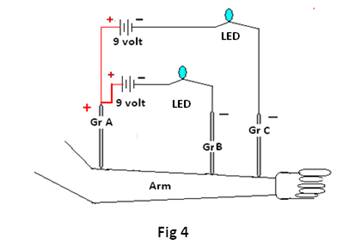
Observations:
i) LEDs do not glow when only GrB & GrC are placed on the skin.
ii) Both the LEDs glow only when GrA (i.e; the common positive electrode) is placed along with GrB &GrC on the skin of the arm.
Discussion:
It appears from the results of experiment 1 that conduction of electrical energy takes place through different modes for different conductors! It is the delocalized electron for the metallic conductor, π electrons for graphite , ions for the distilled water and through depolarization in case of the human body… . as though the membrane is polarized with positive charges outside the membrane (extracellular region) but with negative charges inside the membrane (intracellular region). For cations, the concentration of Na+ ions & K+ ions being 142mEq/L & 4mEq/L respectively in extracellular region, while 14mEq/L & 140mEq/L respectively in intracellular region.
In the case of anions, Cl– ions & HCO3– ions dominate the extracellular region with a concentration of (103mEq/L) &(28mEq/L) respectively, while PO43- and SO42- are found excess in the intracellular fluid with concentration of 75mEq/L & 2mEq/l respectively. This difference in distribution of cations and anions across the cell membrane is believed to generate the normal resting membrane potential of –90millivolts . When both the graphite electrodes are held by hand and the current is said to pass through the human body , more precisely through the nerve the plausible electrical condition inside the nerve is depicted in Fig 5.
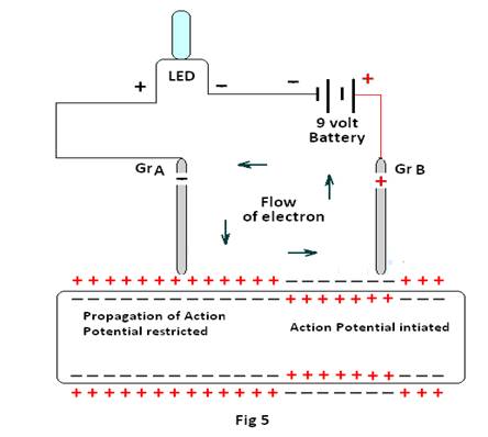
It appears that the positive graphite electrode (Gr B) of the circuit will repel the positive charges outside the membrane and will initiate an action potential as shown in fig 1B. Thereafter, it should propagate as shown in Fig 1D rendering negative charges at the outside of the membrane. On the other hand the negative charge on the graphite electrode (GrA) of the circuit will attract the positive charges on the membrane and hence inhibit cropping up of any negative charges in its vicinity at the outside of the membrane. Thus, any possibility of further propagation of the action potential initiated at Gr B will be restricted. Therefore, how the electrical energy proceeds remains unexplained.
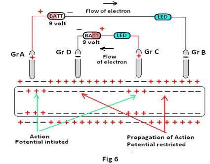
In experiment 2 the case is even more complex, where the propagation of action potential initiated at the positive electrodes of both the circuits seems to be inhibited by the presence of the negative electrode of even one circuit as shown in Fig 6 , i.e., in this case the propagation of action potential initiated at GrA is not only restricted at Gr B but also is restricted at GrD , the negative electrode of the other circuit. But still the LED connected to Gr A & Gr B glows. Thus despite restriction of propagation of action potential at Gr D & Gr B, both the LEDs glow, validating the certain flow of electrons, hence current, in the opposite direction simultaneously. It again shows that more than one electrical impulse can easily be conveyed simultaneously, even in opposite direction, through the human body.
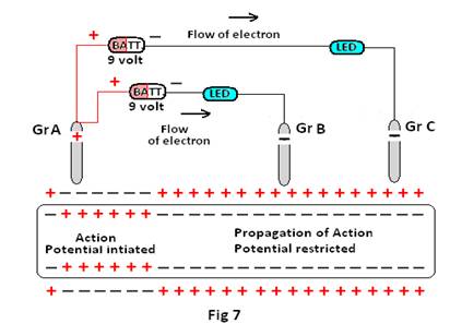
In experiment 3 the case is most interesting, as the propagation of action potential initiated at the common positive electrode (GrA ) is restricted simultaneously by the presence of both the negative electrodes GrB & Gr C of the two circuits (Fig 7). Still, both the LEDs glow simultaneously. It therefore remains a question how electrical impulses from GrA reache at GrC, which is even beyond the negative electrode Gr B of the other circuit.
It is therefore quite clear that the existing theory of nerve conduction (i.e through polarization-depolarization- repolarization ) fails to explain such electrical conduction through nerves.
It can therefore be concluded that as human nerves contain water, their mode of conduction of electrical energy is similar to that of water, 2,3 I.e. orientation of water molecules (the power behind homeopathy) conducts electrical energy in nerves. The cations and anions present in the nerve fiber however help in stabilizing the orientation of water molecules for preferred conduction 4.
Inference :
i) Nerves conduct electrical impulses through orientation of water molecules and the presence of ions only helps in stabilization of the orientations of water molecules.
ii) More than one electrical impulse can easily be conveyed simultaneously, i.e; without any time lag in the same or opposite direction through the human body, hence nerves.
References:
[1] Textbook of Medical Physiology (Eleventh Edition) -Guyton and Hall
[2] The Human Body And Water Both Retain Electrical Energy Ruhul Amin & Biplab Chakraborty (Published in Homeopathy for Everyone Sep2012 hpathy.com
[3]. The key to the Homeopathic dilution, Ruhul Amin & Biplab Chakraborty (Published in Homeopathy 4 Everyone Nov 2012 hpathy.com)
[4] Solutions Ionic or Non-ionic Conduct & Retain Electrical Energy Ruhul Amin & Biplab Chakraborty (Published in Homeopathy for Everyone Jul


One of my patient experienced this type of Electrical nerve shock impulses. The patient was on treatment of Asthma and was given Natrum Mur. 200. The shocks disappeared after completion of treatment. I do not know if Natrum Mur. has anything to do with shock impulses?
What is the principle behind Epilepsy is it the same as explained above if Na and K effects the current flow then is the Epi due to short of the current to the brain nerve due to the deficiency of Na and/or K can NM and phos be administered at the time of Epilepsy the tongue sensory nerves/saliva will carry the deficient Na or K. Pl elaborate This will help Epilepsy patients.
When epilepsy patients get help from homeopathy the reason is that the homeoremedy acts on the brain.