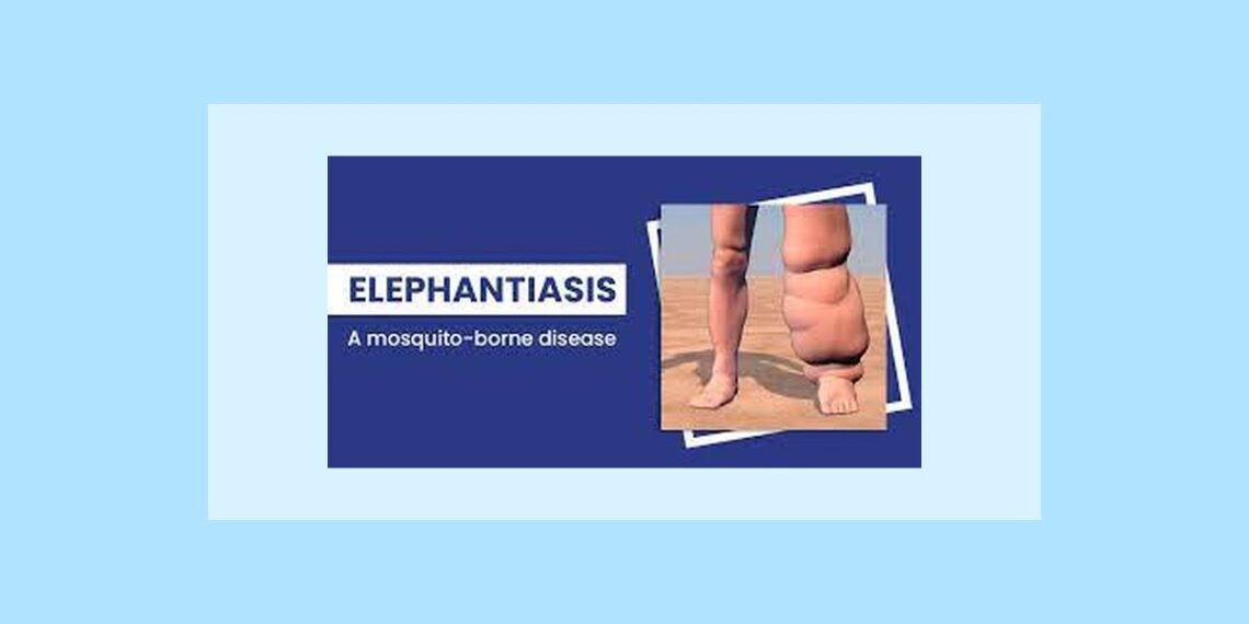
Elephantiasis Massive swelling of the legs also known as lymphatic FILARIASIS, It is caused by obstructed lymph vessels, which prevents drainage of lymph from the surrounding tissue. This causes inflammation and thickening of the vessels walls, eventually blocking them.
More than 120 million people have been affected by elephantiasis: one third in India, another third in Africa, and most of the rest in Southeast Asia, the Pacific, and the Americas. In tropical and subtropical areas where elephantiasis is common, the infection is continuing to increase due to the rapid and unplanned growth of cities, which creates numerous breeding sites for the mosquitoes that transmit the disease.
The parasite worms Wuchereria bancrofti and Brugia malayi cause filariasis, which may lead to elephantiasis. These worms lodge in the lymphatic system (the network of nodes and vessels that maintain the decline fluid balance between the tissues and blood and are an important part of the body’s immune system). The worms live for four to six years, producing millions of immature larvae that circulate in the blood. The infestation spread when mosquitoes bite infected humans and pick up the larvae, which are then passed on into the blood stream of the next human by the bite of the infected mosquito.
Causes of elephantiasis
Elephantiasis is caused by threadlike worms (nematodes) that live in the lymphatic system; it is transmitted by the bite of a mosquito carrying infective larvae. The larvae develop into adult worms in the lymphatic vessels, causing severe damage and swelling (lymphodema).
The most common filarial species is Wuchereria bancrofti, but Brugia malayi and Brugia timori cause the infection in Asia. It takes several mosquito bites over many months to cause the infection. People living in tropical and subtropical settings are at the greatest risk for infection.
Cutaneous filariasis caused by Loa loa is transmitted by the mango fly or deer fly; onchocerca transmits microfilariae via the black fly; and Manosonella streptocerca transmits infection via a midge. Dirofilaria immitis may cause lesions in the lung periphery. Depending on the species, incubation may be 3 to 12 months.
Symptoms and diagnostic path of elephantiasis
Symptoms are similar to human granulocytic ehrlichiosis, including fever, headache, chills, malaise, sweating, muscles aches, nausea, and vomiting. The infection may range from a mild illness to a severe, life threatening disease. It may cause reduction of white blood cells and platelets, anemia, or abnormal liver function.
Differential Diagnosis of Lymph Edema
Cardiac, hepatic, and renal diseases can lead to edema of the lower extremities. They typically cause bilateral leg swelling. Physical examination and laboratory studies are generally able to determine the cause of the disease. Several conditions can coincide. Or clinical importance is differentiation of edema in conjunction with an early stage of venous insufficiency, typically characterized by a pitting edema that decreases or disappears during bed rest, and lipedema.
Post traumatic lymph edema requires differentiation from edema secondary to deep venous thrombosis (DVT) and sympathetic reflex dystrophy syndromes. DVT can be ruled out by duplex sonography, phlebography, contrast enhanced CT venography, or MR venography.
Sympathetic reflex dystrophic burning pain that is not explained by the trauma, hyperpathia, shiny reddish blue discolored skin, and hyperpathia, shiny reddish blue discolored skin, and hyperhidrosis.
X-ray images of the extremities often show signs of de-mineralization in the affected extremity. Differentiation between sympathetic reflex dystrophy syndrome and artificial lymph edema induced by the patient can be difficult and may require surveillance of the patient to rule out self induced strangulation of the extremity.
Lymphostatic elephantiasis (grade III lymph edema) is usually easily diagnosed from the enormous swelling of the affected extremity. It may require differentiation from lipedema, complex angiodysplasia, and other potential causes of elephantiasis such as Recklinghausen’s diseases and lepra tuberose. In contrast to patients with lymph edema, the dorsa of the feet are not usually swollen in patients with lipedema. MRI may help in the differential diagnosis of lymphedema, lipedema, and phlebedma.
Swelling of the upper extremity may require differentiation between Paget -von schroetter syndrome, post thrombotic axillary vein syndrome and lipedema. Patient with paget-van schroetter syndrome typically show spontaneous resolution of the swelling after 4 weeks ultrasound, CT and MRI may be helpful in the assessment of venous patency and can visualize the augmentation of subcutaneous fatty tissue in patient with lipedema.
Prevention of elephantiasis
Controlling the spread and breeding of larvae bearing mosquitoes is the most effective way of preventing elephantiasis. Anyone visiting or living in a danger area, usually in the tropics, should take all the normal precautions, sleeping under mosquito nets and using insect repellent. In general, an American is unlikely to be infested if he or she keeps to hotels and other places that maintain a good standard of cleanliness.
Homeopathic treatment of elephantiasis symptoms– Homeopathy is one of the most popular holistic systems of medicine. The selection of remedy is based upon the theory of individualization and symptoms similarity by using holistic approach. This is the only way through which a state of complete health can be regained by removing all the sign and symptoms from which the patient is suffering. The aim of homeopathy is not only to treat elephantiasis but to address its underlying cause and individual susceptibility. As far as therapeutic medication is concerned, several remedies are available to cure elephantiasis symptoms that can be selected on the basis of cause, sensations and modalities of the complaints. For individualized remedy selection and treatment, the patient should consult a qualified homeopathic doctor in person. There are following remedies which are helpful in the treatment of elephantiasis symptoms:
Apis Mel, Clematis E, Silicea, Natrum Mur, Thuja Occidentalis, Lycopodium, Sulphur, Cannabis Sativa, Aurum Met, Hydrastis and many other medicines.
Reference:
Matthew A. Mauro, Kieran Murphy, Kenneth Thomson, Christoph L. Zollikofer: Image-Guide Intervention; 2008; 1279
Carol Turkington, Bonnie Ashby: The encyclopedia of infectious disease: 2007; 95
David B. Jacoby, R. M. Youngson: Encyclopedia of Family Health; 2004; 554


I think undermentioned medicine are sufficient For Filaria.
Apis Mel, Clematis E, Silicea, Natrum Mur, Thuja Occidentalis, Lycopodium, Sulphur, Cannabis Sativa, Aurum Met, Hydrastis and many other medicines.
I am Thankful you.